


 146 Rue Léo Saignat

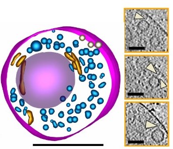
Cryo-correlative light and electron tomography of dopaminergic axonal varicosities reveals non-synaptic modulation of cortico-striatal synapses
Lapios, et al. 2025
Voir la publicationEquipe
David Perrais (team leader, DR CNRS)
Etienne Herzog (PI, DR CNRS)
Julie Angibaud (IE U Bordeaux)
Silvia Sposini (post-doctoral fellow)
Antoine Reynaud (post-doctoral fellow)
Domitille des Robert (PhD student)
Tanuj Arora (PhD student)
Solène Hospital (PhD student)
Mots clés
Vesicular trafficking is one of the most salient features of synaptic physiology. In the tiny (~1 µm wide) chemical synapses, presynaptic vesicles concentrate and release neurotransmitter molecules which bind to post-synaptic receptors. The exocytosis and recycling of synaptic vesicles is a very prominent and essential feature of neuronal physiology that is highly controlled in time and space. Moreover, post-synaptic membrane trafficking, although not as prominent quantitatively, is pivotal for the maintenance of signal transduction complexes supporting synaptic transmission and plasticity. Most of our knowledge about synapse physiology comes from studying glutamatergic synapses which represent the majority of synapses in the brain. Nevertheless, other types of synapses, such as neuromodulatory dopaminergic synapses, could have a very different molecular composition and operate in a different way. However, because they represent a small minority of synapses formed from a very small number of neurons, their analysis has been difficult through classical cellular and molecular methods.
Our goal in the team is to use the most advanced fluorescence imaging techniques together with refined purification of synaptic elements (synaptosomes) to address the mechanisms regulating synapse function through membrane trafficking events in neuronal pre- and post-synaptic compartments. We want to decipher these mechanisms in the normal brain or in the course of disease. To achieve this goal, we use, on top of the standard techniques of the modern neuroscience lab (molecular biology, biochemistry, imaging, electrophysiology), two unique expertise developed by the two PIs: first, with David Perrais, we develop methods to detect individual exocytosis and endocytosis events and perform quantitative imaging. Second, with Etienne Herzog, we purify synaptosomes from adult animals with fluorescence activated synaptosome sorting (FASS), which enables powerful proteomics, transcriptomics and functional approaches. In addition, we have developed, in the last years, fruitful collaborations to analyze the fine structural elements of diverse synapses, in particular neuromodulatory synapses with cryo-correlative light and electron microscopy (cryoCLEM). Cryo-CLEM combined to in-vitro and ex-vivo models of synapses will provide major new insights into the cellular and molecular mechanisms of neurotransmission and neuromodulation.
Altogether, we aim at identifying new pathways in specific synapses and test their relevance for synaptic nanostructure and function in the normal and diseased brain.
Primary neuronal cultures (dissociated, organotypic slices), animals, human induced pluripotent cells differentiated into neurons, cell lines (MEF, NIH 3T3, HEK293)
Lapios, et al. 2025
Voir la publication